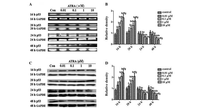
Figure 3.
Expression of p53 in spontaneously differentiating rEHBMCs for a period of two days. Undifferentiated mesenchymal cells express high levels of p53 and a decline was noted during differentiation. (A) and (C) Changes in the levels of p53 in control and ATRA-treated cultures were determined by RT-PCR and western blotting at the indicated time-points. Values shown are representative of at least three independent experiments. GAPDH was used as a loading control. (B) and (D) Densitometric quantification of p53 mRNA and p53 protein were performed. (B) p53 mRNA was subjected to RT-PCR in the exponential growth phase and normalized to GAPDH. aP>0.05, bP<0.05, versus control group; cP>0.05, dP<0.05, versus 0.01 μM ATRA group; eP >0.05, fP<0.05, versus 0.1 μM ATRA group; gP>0.05, versus 1 μM ATRA group. (D) p53 Protein was subjected to western blot analysis in the exponential growth phase and normalized to GAPDH. aP>0.05; bP<0.05, versus control group; cP>0.05; dP<0.05 versus, 0.01 μM ATRA group; eP>0.05, fP<0.05, versus 0.1 μM ATRA group; gP>0.05, versus 1 μM ATRA group. ATRA, all-trans-retinoic acid; rEHBMCs, rat embryo hindlimb bud mesenchymal cells; RT-PCR, reverse transcription polymerase chain reaction.

