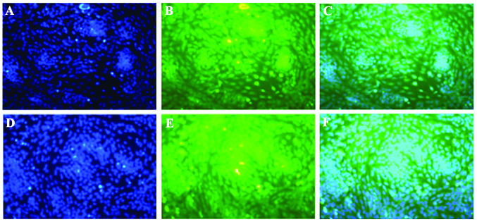
Figure 5.
High levels of (B) p53 and (E) p21 in spontaneously differentiating rat embryo hindlimb bud mesenchymal cells in the absence of any inducing agents at 24 h in vitro. Immunofluorescent microscopy (magnification, ×200) revealed that p53 and p21 were predominantly expressed in the cartilage nodules, mainly in the nucleus. (A) and (D) were stained with DAPI. (C) is a merged image of (A) and (B) and (F) is a merged image of (D) and (E).

