Figure 3.
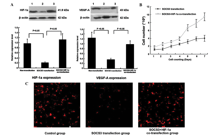
HIF-1α is required for proliferation of SOCS3-transduced SCLC cells and their angiogenic potential. (A) Western blot analysis of HIF-1α and VEGF-A protein expression. Representative images of three independent experiments (lane 1, HIF-1α and VEGF-A protein expression in the non-transfection group; lane 2, VEGF-A and HIF-1α protein expression in SOCS3 transfection group; lane 3, HIF-1α and VEGF-A protein expression in SOCS3 and HIF-1α co-transfection group) and quantified expression of HIF-1α and VEGF-A protein normalized to β-actin (HIF-1α expression in SOCS3 transfection group vs. non-transfection group, P<0.05; HIF-1α expression in SOCS3 transfection group vs. SOCS3+HIF-1α co-transfection group, P<0.05; VEGF-A expression in SOCS3 transfection group vs. non-transfection group, P<0.05; VEGF-A expression in SOCS3 transfection group vs. SOCS3+HIF-1α co-transfection group, P<0.05) (B) Growth curves of cells in SOCS3 transfection and SOCS3+HIF-1α co-transfection groups. Co-transfection with SOCS3 and HIF-1α significantly promoted the growth rate of the cells (NCI-H446/SOCS3 group vs. NCI-H446/SOCS3+HIF-1α group, P<0.05). (C) CD34 expression in the three groups (magnification, ×200). Immunofluorescence staining showed that following transfection with SOCS3, CD34 expression intensity was significantly decreased, while it was significantly intensified following upregulation of SOCS3 and HIF-1α. SOCS3, suppressor of cytokine signaling 3; VEGF, vascular endothelial growth factor; SCLC, small cell lung cancer; HIF-1α, hypoxia-inducible factor-1α.

