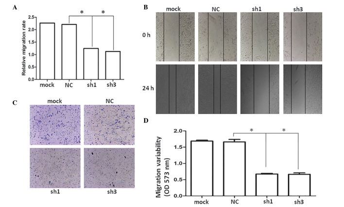
Figure 4.
CCL2 promotes LM8 cells invasion in vitro. (A) Wound-healing assay results. (B) Five representative images of the scratched areas were captured under a light microscope at 0 and 24 h. The average gap of the time-points was used to quantify the wound healing ability of each group (magnification, ×50). (C) Transwell assay of the LM8, NC, LM8-sh1 and LM8-sh3 cells (magnification, ×100; crystal violet stain). (D) Quantification of migration variability. *P<0.05, compared with NC. mock, untransfected; NC, negative control; sh, short hairpin.

