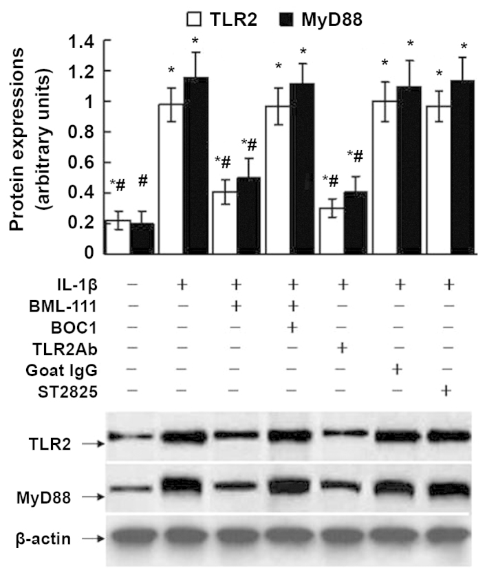
Figure 9.

Expression of TLR2 and MyD88 assessed using western blot analysis of leukocytes exposed to IL-1β. The cultured leukocytes were obtained from a normal control mouse and stimulated with IL-1β (10 ng/ml) for 30 min with or without pre-treatment of BML-111 (1 mM) for 30 min, antagonist of G protein-coupled LXA4 receptor BOC1 (100 μM) for 30 min, TLR2Ab (1 μg/ml) for 1 h, goat IgG (1 μg/ml) for 1 h and MyD88 dimerization inhibitor ST2825 (20 μM) for 1 h. The western blot is representative of five independent experiments, and the lower panel shows β-actin protein, which served as a loading control. Semiquantitative analysis was performed by using UVP-gel densitometry. Arbitrary unit = (ATLR2/Aβ-actin) ×100%, or (AMyD88/Aβ-actin) ×100%. Values are expressed as the mean ± standard deviation of five independent experiments. *P<0.05, as compared to the arbitrary units of the same protein in the cells without treatment. #P<0.05, as compared to the arbitrary units of the same protein in the cells treated with IL-1β alone. IL, interleukin; IgG, immunoglobulin; TLR, toll-like receptor; TLR2Ab, toll-like receptor 2-neutralizing antibody; BOC1, N-t-Boc-Phe-Leu-Phe-Leu-Phe.
