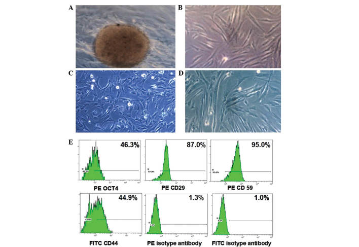
Figure 1.
Cellular characteristics of HUMSCs. (A) HUMSCs were observed to migrate out of the tissue fragment and exhibited a fibroblast-like morphology at passages (B) 1, (C) 3 and (D) 9. (E) Flow cytometric analysis of HUMSCs using antibodies against the human stem cell markers octamer-binding transcription factor 4, CD29, CD44 and CD59. PE- or FITC-labeled isotype antibodies were used as controls. Magnification, ×100. HUMSCs, human umbilical cord mesenchymal stem cells; PE, phycoerythrin; FITC, fluorescein isothiocyanate.

