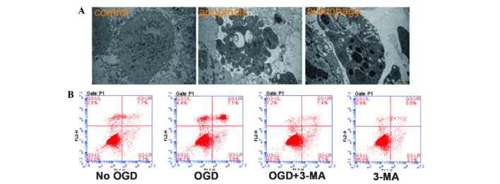
Figure 2.

Hypoxia-induced apoptosis and autophagy in HUVECs. (A) HUVECs were treated with OGD for 4 h followed by 12 h reoxygenation. Apoptosis and autophagy were then observed using electron microscopy (magnification, ×12,000). (B) HUVECs were treated with 3-MA and apoptosis was measured using flow cytometry. Early apoptotic or apoptotic and necrotic cells were identified as single positive for FITC-Annexin V (lower right quadrant) or double positive for both FITC-Annexin V and propidium iodide (upper right quadrant), respectively. HUVECs, human umbilical vein endothelial cells; OGD, oxygen-glucose deprivation; FITC, fluorescein isothiocyanate; 3-MA, 3-methyladenine.
