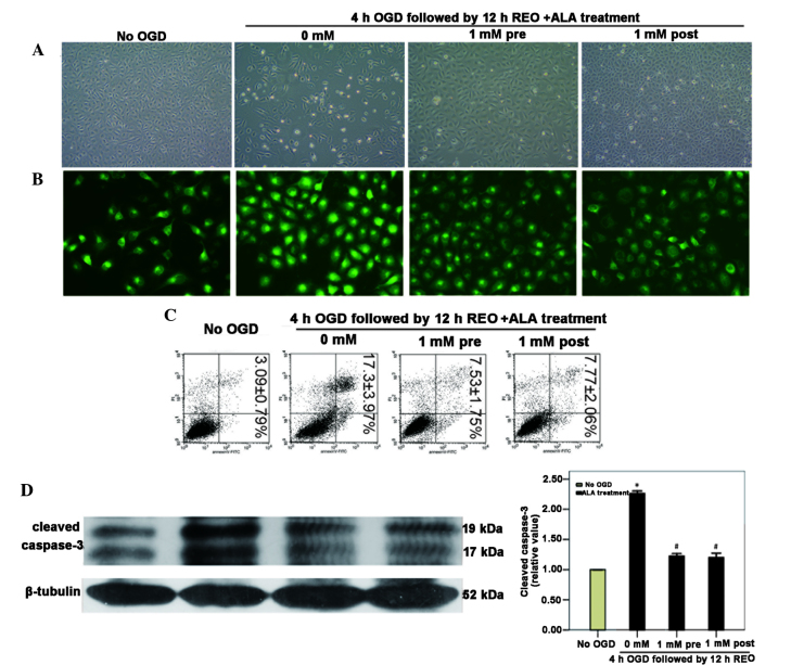
Figure 3.
ALA pre- or post-treatment reduces HUVEC apoptosis induced by OGD/reoxygenation. HUVECs were subjected to 4 h of OGD followed by 12 h of reoxygenation in the presence or absence of 1 mM ALA pre- or post-treatment. (A) Cell morphology was observed using inverted phase contrast microscopy (magnification, ×100). (B) Fluorescence microscopy with Rho123 staining was used to detect the mitochondrial membrane potential (magnification, ×400). (C) Cell apoptosis was measured using flow cytometry. Percentages of apoptotic cells (lower right quadrant) as well as apoptotic and necrotic cells (upper right quadrant) are presented as the mean ± standard deviation (n=3). (D) Western blot analysis was used to measure cleaved caspase-3 expression levels and quantitative analysis of these western blots revealed that cleaved caspase-3 was significantly downregulated in ALA pre- or post-treatment groups. *P<0.05 vs. no OGD; #P<0.05 vs. 0 mM ALA treatment. HUVECs, human umbilical vein endothelial cells; OGD, oxygen-glucose deprivation; ALA, α-lipoic acid; REO, reoxygenation.

