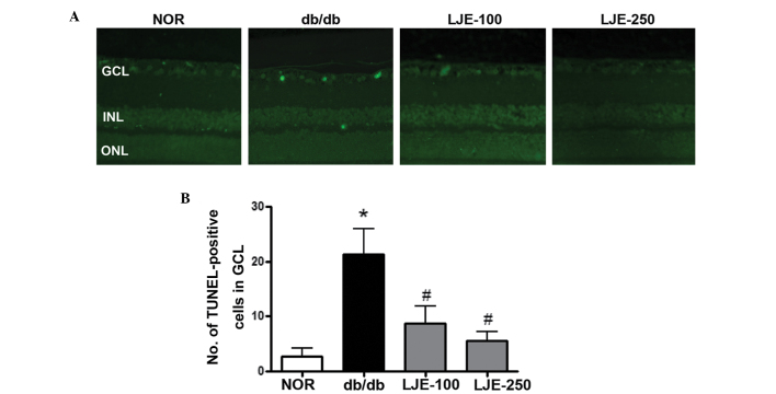
Figure 3.
Apoptosis of retinal ganglion cells. (A) Retinal sections were stained with TUNEL (green). Apoptotic ganglion cells were observed in the vehicle-treated db/db mice (magnification, ×200). db/db, diabetic db/db mice; LJE-100, db/db mice treated with LJE (100 mg/kg); LJE-250, db/db mice treated with LJE (250 mg/kg). (B) Quantitative analysis of TUNEL-positive cells in GCL. Values are expressed as the mean ± standard error of the mean, n=8. *P<0.01, vs. NOR mice, #P<0.01, vs. vehicle-treated db/db mice. TUNEL, terminal deoxynucleotidyl transferase dUTP nick end labeling; GCL, ganglion cell layer; INL, inner nuclear layer; ONL, outer nuclear layer; NOR, normal; LJE, Litsea japonica extract.

