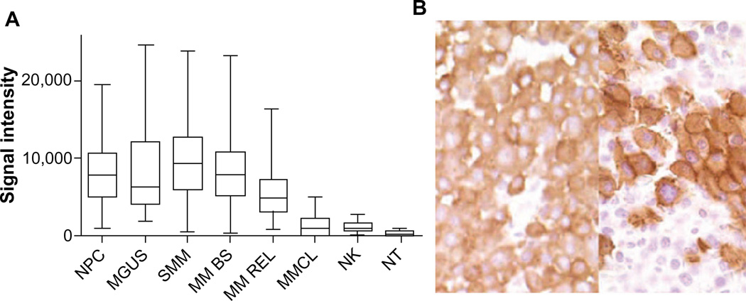
Figure 1.
CS1 expression quantified by gene expression (A), and immunohistochemistry (B).
Notes: (A) CS1 is highly expressed in CD138+ NPC (n=24), patients with MGUS (n=14), SMM (n=35), and MM BS (n=532) and MM REL (n=79), while expression in MMCL (n=45) is lower. CS1 is expressed in NK cells but at a lower level (NK, n=16), and is nearly negligible in other NT (15 tissue samples). (B) Strong CS1 expression in plasma cells from BM samples of two patients with MM by immunohistochemical staining with mAb IG9 (400×). Images shown in (B) are reproduced from Hsi et al. CS1, a potential new therapeutic antibody target for the treatment of multiple myeloma. Clin Cancer Res. 2008;14(9):2775–2784.18 Copyright © 2008, American Association for Cancer Research.
Abbreviations: mAb, monoclonal antibody; MGUS, monoclonal gammopathy of undetermined significance; MM BS, multiple myeloma treated on Total Therapy 2 and 3 protocols at baseline; MM REL, multiple myeloma treated on Total Therapy 2 and 3 protocols at relapse; MM, multiple myeloma; MMCL, myeloma cell lines; NK, natural killer; NPC, normal plasma cells from healthy donors; NT, normal healthy tissue; SMM, smoldering multiple myeloma.

