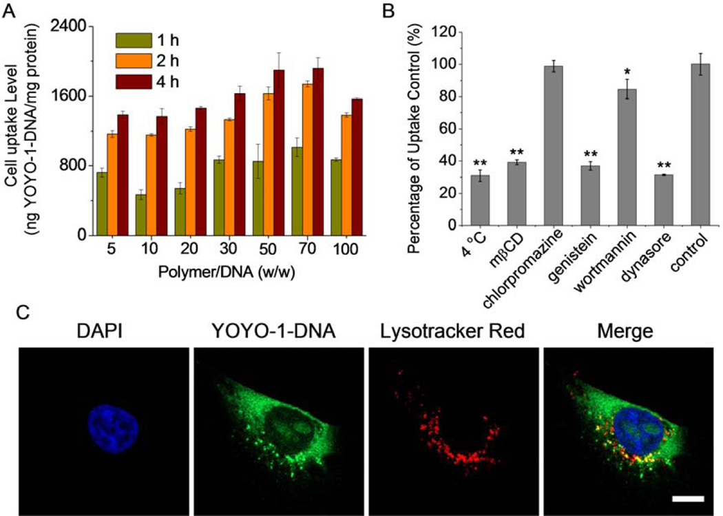
Fig. 6.
Intracellular kinetics of P1–13700/YOYO-1-DNA polyplexes (weight ratio of 50) in HeLa cells. (A) Uptake level of polyplexes following incubation at 37 °C for different time (n = 3). (B) Uptake level of polyplexes at 4 °C or in the presence of various en docytic inhibitors. Results were represented as percentage (%) of the uptake level at 37 °C and in the absence of inhibitors (n = 3). (C) CLSM images showing the cellular internalization and distribution of polyplexes in HeLa cells following incubation at 37 °C for 4 h. Cell nuclei were stained with DAPI and the endosomes/lysosomes were stained with Lysotracker Red. Bar represented 10 µm.

