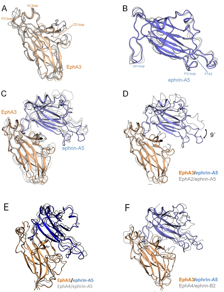
Fig 3. Ephrin-A5 is markedly tilted when bound to EphA3.
(A) The ephrin-A5-bound EphA3 LBD (colored wheat) superimposes well to the EphA LBDs from other EphA/ephrin-A structures (depicted as grey lines; 0.5–0.7 Å RMSD to PDBIDs 3MX0 (EphA2/ephrin-A5), 3HEI (EphA2/ephrin-A1), and 2WO3 (EphA4/ephrin-A2)). (B) Ephrin-A5 (colored light blue) superimposes well to previously available ephrin-A5 structures (0.5–0.6 Å RMSD to PDBIDs 1SHX (unbound ephrin-A5), 1SHW (EphB2/ephrin-A5), 3MX0 (EphA2/ephrin-A5), and 2X11 (EphA2/ephrin-A5)). (C) The EphA3/ephrin-A5 complex superimposes less well to other EphA/ephrin-A complexes (1.7–2.1 Å RMSD compared to 3MX0, 2X11, 2WO3 and 3HEI). (D) Superposition of the EphA2/ephrin-A5 complex (PDBID 3MX0) to just the EphA3 LBD reveals that ephrin-A5 is tilted by 9° in the EphA3/ephrin-A5 structure compared to its orientation in complex with EphA2. This is because the entire ephrin-A5 molecule, including the GHL loop, is tilted. (E) The EphA3/ephrin-A5 complex is oriented similarly to an EphA4/ephrin-A5 complex (PDBID 4M4R) that also shows a similar tilt of ephrin-A5 (Table 3). (F) The EphA3/ephrin-A5 complex is oriented similarly to the EphA4/ephrin-B2 complex (PDBID 2WO2).

