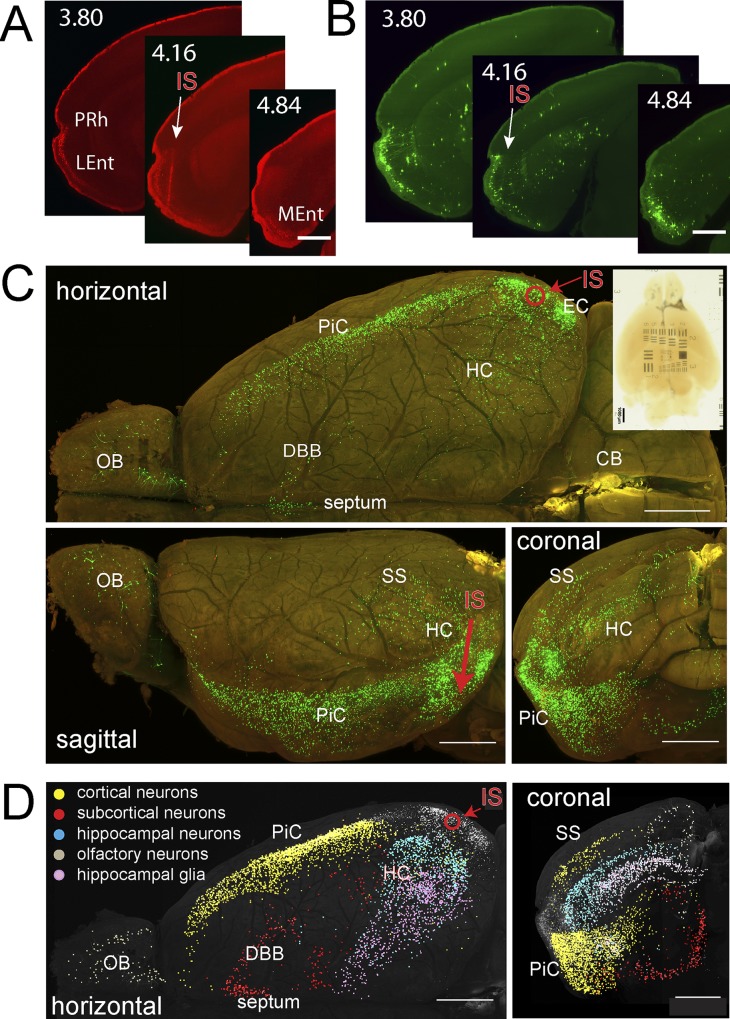
Fig 5. Visualization of neurons monosynaptically connected to RABVΔG-EGFP(EnvA) infected neurons in EC.
The fixed brain was cleared with tB-BABB/30°C for 64d. Red fluorescence (rAAV- mRFP1-IRES-TVA) and green fluorescence (RABVΔG-EGFP) were recorded with the LSFM(xy pixel size, 1.61 μm; z-step size, 3.23 μm). Maximum projections were generated after correction for tissue-generated signal attenuation. (A, B) Maximum projections (50 μm) from coronal re-slices of the horizontal recording of red fluorescence (A, rAAV- mRFP1-IRES-TVA) and green fluorescence (B, RABVΔG-EGFP). The precise position of each projection is given as distance to bregma according to [44]. Injection site and direction are indicated by a white arrow. Prh, Perirhinal Cortex; LEnt, Lateral Entorhinal Cortex; MEnt, Medial Entorhinal Cortex; IS, injection site. Scale bar, 0.5 mm. (C) Red/green overlays of maximum intensity projections generated from z stacks of stitched xy horizontal mosaic planes (top), and from sagittal (bottom left) and coronal re-slices (bottom right) of these z stacks. Bright green structures show RABVΔG-EGFP-positive cells. The red circle and the red arrow indicate the positions of the dorsoventral virus injection channel in horizontal and sagittal views, respectively. The transmission image of the whole cleared brain located on top of a translucent USAF 1951 resolution target is shown in the upper right corner. Scale bars, 0.5 mm. (D) Maximum projections (left, horizontal; right, coronal) of a KNOSSOS 3D skeleton dataset generated from the green channel dataset. Each dot indicates the position of an EGFP-labeled, manually marked cell. Color coding: grey, cells in injection region (EC area); marked cells located outside the injection area are color coded as indicated. Respective greyscale maximum projections of the green channel fluorescence were overlaid to depict tissue location. OB, Olfactory bulb; DBB, Diagonal Band of Broca; PiC, Piriform Cortex; SS, Somatosensory Cortex; HC, Hippocampus; EC, Entorhinal Cortex; IS, injection site. Scale bars, 0.5 mm.

