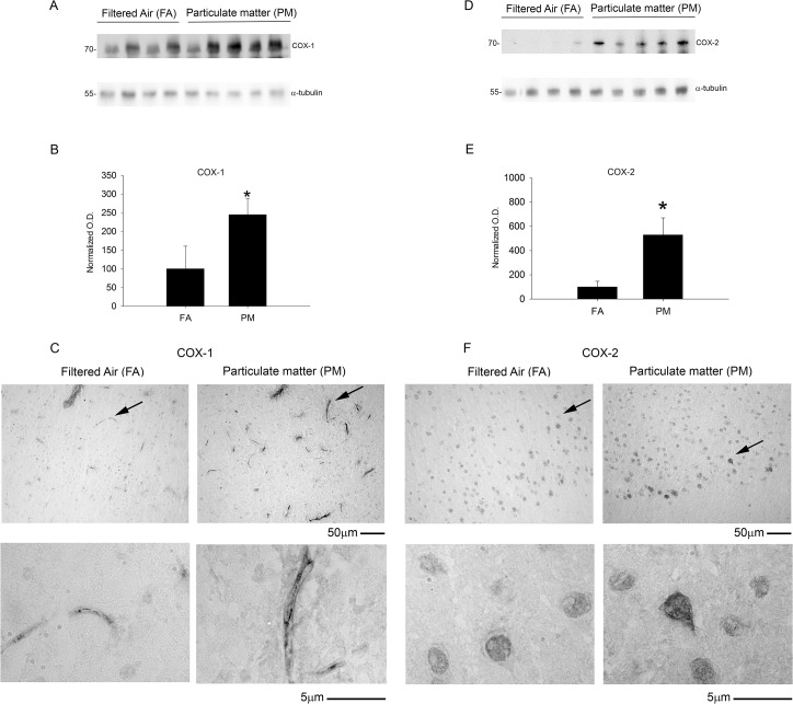
Fig 9. Brain cyclooxygenase enzyme (COX-1 and COX-2) protein levels increased with 9 month airborne particulate matter exposure.
Brains of 9 month exposed filtered air (FA) or PM2.5 (PM) mice were collected with the left hemisphere fixed for immunohistochemistry and the right temporal cortices collected for biochemical analysis. Temporal cortex lysates from FA and PM brains were used for western blot analysis using A, anti-COX-1, D, anti-COX-2 and α-tubulin (loading control) antibodies. Optical densities for B, COX-1 and E, COX-2 were normalized to their respective loading controls, averaged (+/-SD), and graphed, *p<0.05. Brains were immunostained using C, anti-COX-1 and F, anti-COX-2 antibodies. Images shown are representative from the temporal cortex. Arrows indicate regions taken for higher magnification.

