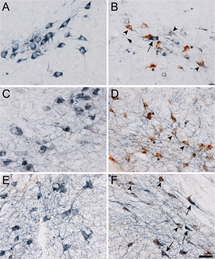
Fig 4. Upregulation of Sprr1a, Gadd45a and Sox11 early after intrastriatal 6-OHDA lesion occurs specifically in substantia nigra (SN) dopamine (DA) neurons. A-F).
The mRNA for Sprr1a (A and B), Gadd45a (C and D) and Sox11 (E and F) was identified using RNAscope in situ hybridization (ISH; brown puncta) and DA neurons in the SN were identified with immunohistochemistry for tyrosine hydroxylase (blue staining, a DA neuron marker) in animals one week after an intrastriatal 6-OHDA lesion. The DA neurons on the intact side (i.e. contralateral to the 6-OHDA lesion) had little to no ISH signal due to the normally low basal expression of these RAGs (A, C, and E). In contrast, the DA neurons on the lesioned side had robust ISH signal (B, D and F). In the lesioned side, DA neurons appeared unhealthy and three cellular phenotypes were present, 1) little to no RAG ISH but strong TH indicating an apparently healthy neuron (arrow), 2) mild-moderate RAG ISH with moderate-weak TH staining indicating a DA neuron that is likely enroute to degenerating (arrowhead), and 3) robust ISH signal and little-no TH staining likely indicating DA neurons closer to degenerating (asterisk; DA neuron determination is based on neuroanatomical location within the SN and cell morphology). These data indicate that the upregulation of Sprr1a, Gadd45a and Sox11 identified by gene arrays and confirmed by qPCR occurred specifically in SN DA neurons. Scale bar = 50μm.

