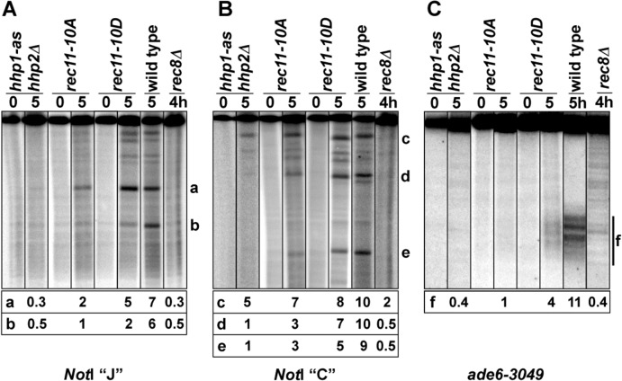
Fig 1. Meiotic DNA breakage in hhp, rec11, and rec8 mutants.
Strains with the indicated mutations were induced for meiosis in the absence of an ATP analog. At the indicated times, DNA was extracted and analyzed by Southern blot hybridization. All time points for each mutant were run on the same gel, one gel for the two NotI fragments and another for the ade6-3049 fragment (S1 Fig). Data below each lane with meiotically induced DNA are the percent of total DNA in the bands labeled a—f after subtraction of the intensity in the corresponding 0 hr lane. These data reflect DNA breakage at DSB hotspots. DSBs at the ade6-3049 hotspot are spread over about 1 kb, indicated by the bar (f) on the right. See also S1 Fig. (A) The 501 kb NotI fragment J on chromosome 1 was analyzed with a probe at its left end [60]. (B) The 1.5 Mb NotI fragment C on chromosome 2 was analyzed with a probe near its left end [18]. (C) The 6.6 kb AflII fragment containing ade6 on chromosome 3 was analyzed with a probe at its right end [61].

