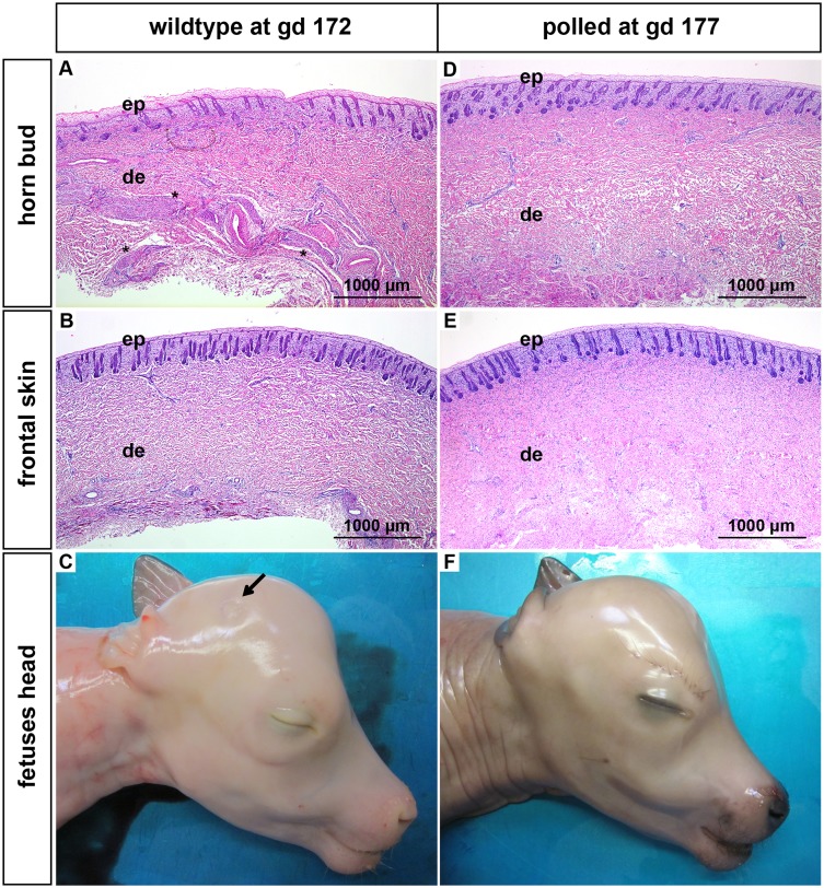
Fig 6. Features of horn buds and frontal skin from wild type and polled fetuses (gd 172 and 177).
(A): Horn bud from a wild type fetus with multiple layers of vacuolated keratinocytes. Note presence of thick nerve bundles in the dermis below the horn bud (black stars). (B): Frontal skin from a wild type fetus. Note absence of thick nerve bundles in the dermis. (C): Macroscopic picture of a horn bud from a wild type fetus. Note indentation of skin (black arrow). Region of the horn bud (D) and frontal skin (E) from a polled fetus. Note absence of thick nerve bundles in the dermis. (F): Macroscopic picture of a polled fetus without indentation of the skin. Haematoxylin and eosin. Gd = gestation days, ep = epidermis, de = dermis.

