
Figure 2.
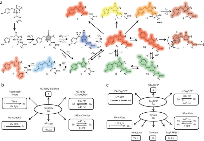
Major chemical transformations of the chromophores in red fluorescent proteins. (a–c) Transformations in fluorescent protein subfamilies derived from red fluorescent protein (a), mCherry (b) and TagRFP (c). The colored shading of the chemical structures (a) and chromophore numbers (b,c) correspond to the spectral range of the chromophore fluorescence emission. Gray shading denotes the nonfluorescent state; [H] denotes reduction; and [O] denotes oxidation. The chromo states (structures 5, 10 and 13) are not necessarily caused by a cis-trans chromophore isomerization but may result from modifications of the chromophore environment of the same isoform that decrease quantum yield. hv, photon.