
Figure 1.
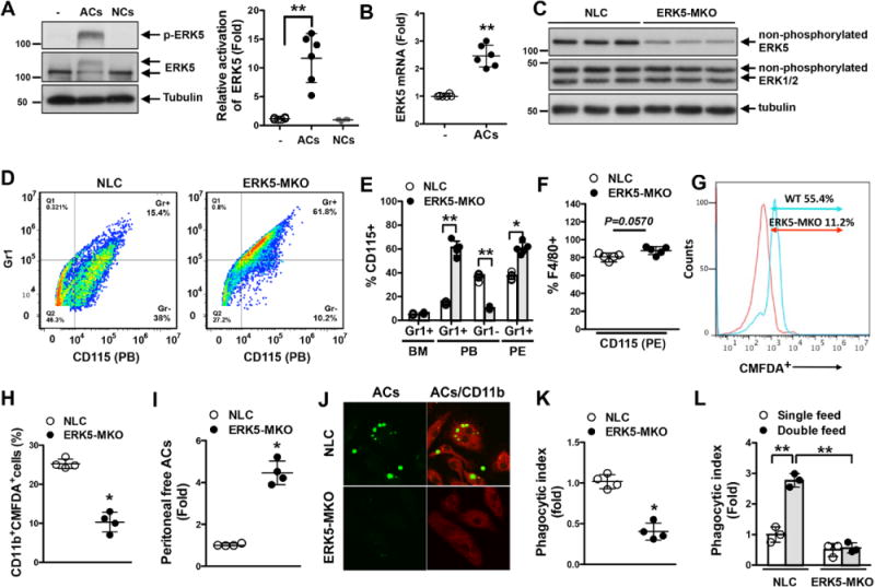
ERK5 kinase activation is involved in macrophage efferocytosis. A and B, ERK5 phosphorylation (A) and mRNA expression (B) in bone marrow–derived macrophages fed apoptotic cells (ACs) for 30 minutes (A) or 18 hours (B), respectively. Feeding necrotic cells (NCs) did not increase ERK5 phosphorylation (A, left), and the graph represents densitometry data (A, right). − indicates no treatment. Data are mean±SD (n=6); **P<0.01. C, ERK5 expression in peritoneal macrophages isolated from nontransgenic littermate control (NLC; LysCre+/−) and LysCre+/−ERK5fl/fl (ERK5-MKO) mice. Cell lysates were immunoblotted for total ERK5, ERK1/2, and tubulin. n=3 per genotype. D through F, Characterization of CD115+ and Gr1+ cells identified by specific immunolabeling. D and E, Representative flow cytometry results showing CD115+Gr1+/− cells in peripheral blood (PB; D) and quantified data for bone marrow (BM), peripheral blood, and peritoneal (PE; E) cells from NLC or ERK5-MKO mice. F, Maturation levels of peritoneal macrophages from NLC and ERK5-MKO mice identified by the expression of CD115 and F4/80 markers. G, Representative flow cytometery data on AC phagocytosis by NLC and ERK-MKO macrophages in vitro after 60 minutes of incubation with 5-chloromethylfluorescein diacetate (CMFDA)–labeled ACs at a 1:5 (bone marrow–derived macrophages:ACs) ratio (n=5). H through K, In vivo clearance and phagocytosis assays. H, CMFDA-labeled ACs were injected intravenously, and 12 hours later, splenic macrophages (CD11b+) that have ingested labeled ACs were quantified by flow cytometery. I, CMFDA-labeled ACs were injected into the peritoneum of NLC and ERK5-MKO mice, and 6 hours later, free fluorescent cells in the peritoneal cavity were quantified. J, Representative images showing phagocytized and free ACs. Anti-CD-11b staining (red) was used to identify macrophages. Delayed clearance of ACs by resident peritoneal macrophages in ERK5-MKO mice is illustrated. K, Phagocytic index 6 hours after AC injection. See Methods for details. H, I, and K, Data are mean±SD (n=4); *P<0.05. L, Bone marrow–derived macrophages isolated from NLC and ERK5-MKO mice were fed ACs 24 hours before refeeding them with CMFDA-labeled ACs. Macrophages with engulfed fluorescent ACs were quantified by flow cytometery 24 hours later. The phagocytic index was determined as described in Methods. Data are mean±SD (n=4); **P<0.01. Comparison between 2 independent groups was subjected to Wilcoxon rank sum test.