
Figure 6.
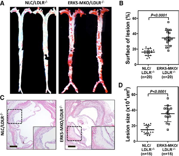
Accelerated atherosclerotic plaque formation in ERK-MKO/low-density lipoprotein receptor (LDLR)−/− mice. A, Whole aortas from each mouse group were stained by Oil-red O. C, Sections from the proximal aortas of each group were stained by hematolylin-eosin. Bars, 10 μm. B and D, The area covered by Oil-red O staining is expressed as percentage of the total surface area of aorta from nontransgenic littermate control (NLC)/LDLR−/− and ERK5-MKO/LDLR−/− mice (B). The area of atherosclerotic plaques in sections of the proximal aorta was measured (D). Data are mean±SD (B, n=20; D, n=15 per genotype); **P<0.01. Comparison between 2 independent groups was subjected to Wilcoxon rank sum test.