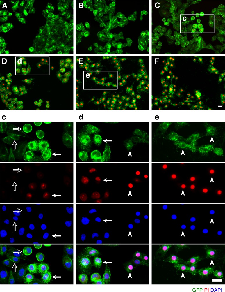
Fig 2. ETX induces rapid internalization of rMAL that precedes or coincides with cell death.
CHO cells stably expressing GFP-rMAL (green) were treated with 50 nM ETX in the presence of 2 μg/ml PI (red) and fixed at 0 minutes (A), 5 minutes (B), 15 minutes (C), 30 minutes (D), 60 minutes (E), and 90 minutes (F) following the treatment. Regions (c-e) framed in C-E are shown at a higher magnification to illustrate morphological details. Images in c-e are shown in separate fluorescence channels and as an overlay plus a DAPI-counterstain. Hollow arrows in c point to the cells on which MAL is largely present on the cell surface. Noticeably, these cells are negative for PI staining. The morphological heterogeneities likely reflect different phases of cell cycle in individual cells within the population. Arrows in c and d point to the cells displaying intracellular vesicles of MAL protein, which indicate an ongoing MAL internalization process in response to ETX. These are dying cells that exhibit PI inclusion in their nuclei with a higher staining density in nucleoli. Arrowheads in d and e point to the dead cells that display condensed nuclei brightly stained with PI and contain intracellular MAL protein. Scale bars represent 20 μm. Data shown are representative of at least three independent experiments.

