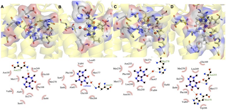
Fig 2. CFF’s most populated binding poses in systems I-III (A-D).
For each binding pose, the upper panel shows the protein backbone in yellow cartoon, CFF and residues interacting with CFF in thick and thin sticks, respectively. Water molecules forming H-bonds with CFF and residues are represented as red sphere; the lower panel shows the corresponding 2-d chart.

