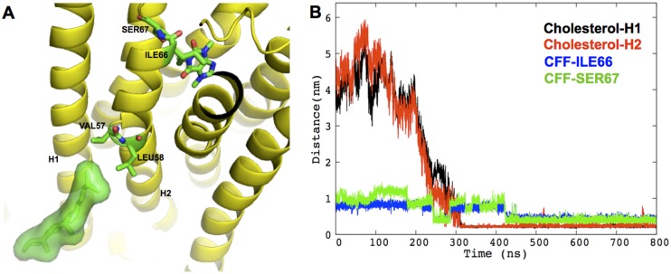
Fig 3. Specific cholesterol binding to hA2AR A) Cartoon showing cholesterol-binding pose in H1/H2 cleft in system III.

The receptor is shown in yellow cartoon; the cholesterol molecule is shown as green sticks surrounded by its solvent accessible surface; CFF, cholesterol-interacting residues, VAL57, LEU58, as well as CFF-interacting residues ILE66, SER67 are shown as green sticks with oxygen and nitrogen atoms colored in red and blue, respectively. B) The diffusion of cholesterol into of the H1/H2 cleft enhances hydrophobic contacts between CFF and H2. The minimum distances between the specific cholesterol molecule and H1 (residues 5–34), between cholesterol and H2 (residues 41–67), between C5@CFF and heavy atoms of ILE66 and SER67 side chains, are shown in black, red, blue and green, respectively.
