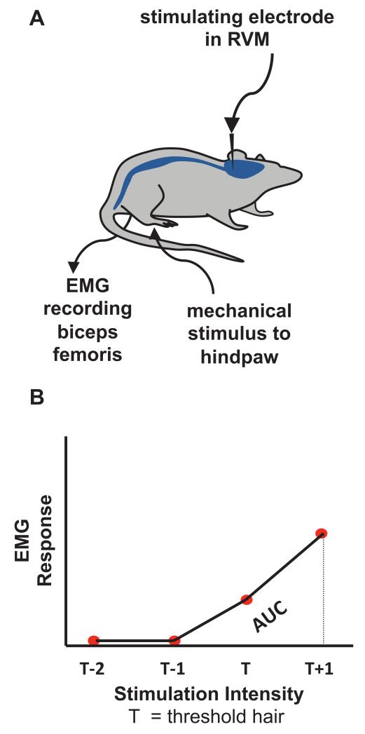
Fig. 1.
(A) Schematic diagram illustrating the experimental set-up. A stimulating electrode was placed in the rostroventral medulla (RVM) of lightly anaesthetised rats, with electromyography (EMG) recording electrodes inserted into biceps femoris. Before and during RVM stimulation, hindpaws were mechanically stimulated with von Frey hairs and evoked EMG responses were recorded. (B) Quantification of overall reflex response. Von Frey hairs of increasing intensity were applied until an increase in EMG activity 10% greater than background was evoked. This hair was designated the threshold hair (T). EMG responses to two subthreshold (T-2 and T-1) and one suprathreshold hair (T+1) were quantified from the average of three applications and plotted against stimulus intensity to generate a baseline stimulus-response profile. The area under this curve (AUC) was then calculated to quantify overall reflex response and spinal excitability.

