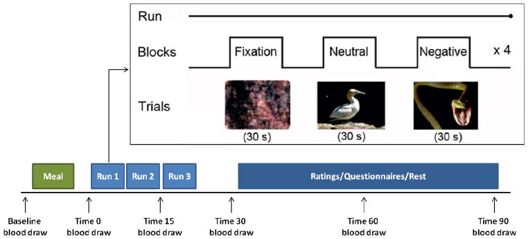
Figure 1. Illustrative Schemata of the fMRI Stress Response Task* and Timed Blood Draws.

* fMRI task adapted from International Affective Picture System (Goldstein et al, 2005b; Lang et al, 2008; Goldstein et al, 2010b). Baseline blood draw was acquired at ~8am, fasting since midnight. A baseline in-scanner blood was acquired and then draws were timed to hormonal response to stress, i.e., pituitary (15, 30 min. post-stress challenge) and steroid hormones (60, 90 min. post-stress challenge).
