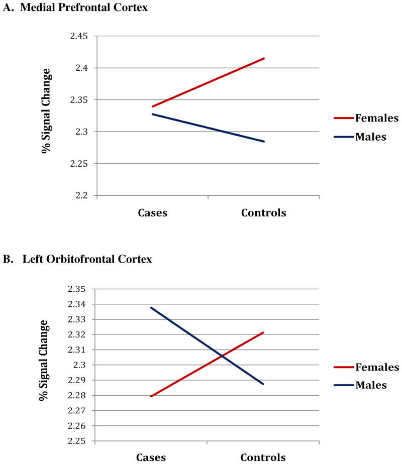
FIGURE 4. Interaction of Sex by Case Status on Signal Intensity Changes in Prefrontal Cortices at High Levels of Cortisol:DHEAS (i.e., hypercortisolemia).
Graphs represent mean percent signal change (natural log transformed BOLD) in the medial prefrontal cortex (A) and left orbitofrontal cortex (B) for subjects in the highest 75th percentile of the healthy control cortisol:DHEAS distribution at time 90. Hypercortisolemia in male cases was associated with higher medial prefontal and left orbitofrontal cortex signal changes compared with healthy controls and lower activity for female cases compared with healthy controls

