
Fig. 1.
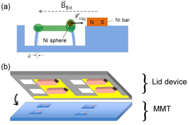
(a) Schematic of magnetic microtissue actuation system, showing a microtissue (green) whose tension deflects flexible micropillars. Application of a magnetic field BExt magnetizes the 100 μm diameter Ni sphere mounted on one of the micropillars, and also a nearby small Ni bar, creating a force between the sphere and the bar, FMag, which stretches the microtissue. The deflection δ of the left pillar reports the tissue’s force. (b) Schematic of a Si wafer “lid” device, showing Au fingers that serve as electrodes for electrodeposition of Ni bars (tan shapes) and holes (white squares) etched through the wafer that allow optical and media access to the microtissues. Alignment with the microtissue array is as indicated.