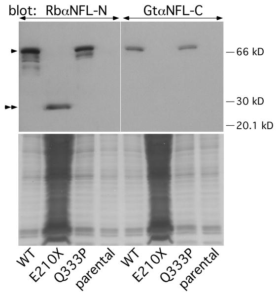
Fig. 6. Detection of the E210X mutant protein by immunoblotting.
The upper panel shows immunoblots of lysates from parental SW-13 vim- cells or cells transiently transfected to express WT human NFL (WT), E210X, or Q333P, probed with antisera against the N-terminus (left panel) or C-terminus (right panel) of NFL. Each lane contains 60 μg of lysate, except for the E210X sample, which was deliberately overloaded (∼1800 μg) to show the fainter NFL band (double arrowheads) of the truncated E210X mutant. The positions of the 20, 30, and 66 kDa size markers are shown, and full-length NFL protein is indicated by the single arrowhead. The lower panels are images of the Coomassie-stained gels after transfer.

