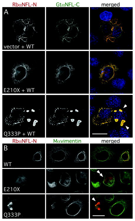
Fig. 7. The E210X mutant does not affect the ability of WT NFL or vimentin to form a network.
(A) These are deconvolved images of SW-13 vim- cells, transiently transfected to express WT human NFL (WT) plus “empty vector” (vector), E210X, or Q333P, then immunostained with a rabbit antiserum against the N-terminus of NFL (red), a goat antiserum against the C-terminus of NFL (green), a mouse monoclonal antibody against vimentin (not shown), and counterstained with DAPI (blue). Note the filamentous network of NFL staining in cells expressing WT NFL plus vector or E210X, whereas cells expressing both WT NFL and Q333P form cytoplasmic aggregates (arrowheads). Scale bar: 20 μm. (B) These are deconvolved images of SW-13 vim+ cells, transiently transfected to express WT human NFL (WT), E210X, or Q333P, then immunostained with a rabbit antiserum against the N-terminus of NFL (red) and a mouse monoclonal antibody against vimentin (green). Note that vimentin and NFL staining are co-localized in cells expressing WT NFL (forming a network) or Q333P (which collapses the network and forms cytoplasmic aggregates; arrowheads), but not in cells expressing E210X, which is found in small puncta/diffuse staining that did not disrupt vimentin assembly (double arrowheads). Scale bar: 20 μm.

