Figure 2.

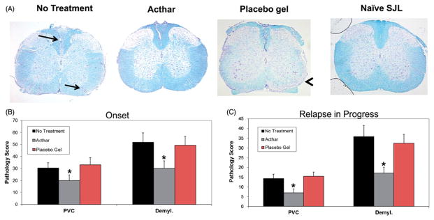
Acthar treatment protects EAE mice from inflammation and demyelination. (A) Representative Luxol fast blue-stained sections of mouse spinal cords. Arrows perivascular cuffs; Arrowhead: demyelination. Images shown at 4× magnification. (B) Pathology scoring of spinal cord tissue sections obtained from mice in which treatment was initiated at the onset of relapse (n = 15 for no treatment, 12 for Acthar treatment, 10 for Placebo gel treatment). (C) Pathology scoring of spinal cord tissue sections obtained from mice in which treatment was initiated after the onset of relapse (n = 16 for all groups of mice). Pathology scoring was performed as described in the “Methods” section. Values represent the mean ± SEM (at least six spinal cord tissue sections were scored for each mouse). PVC perivascular cuffing; Demyl. demyelination. *p<0.05, t test.
