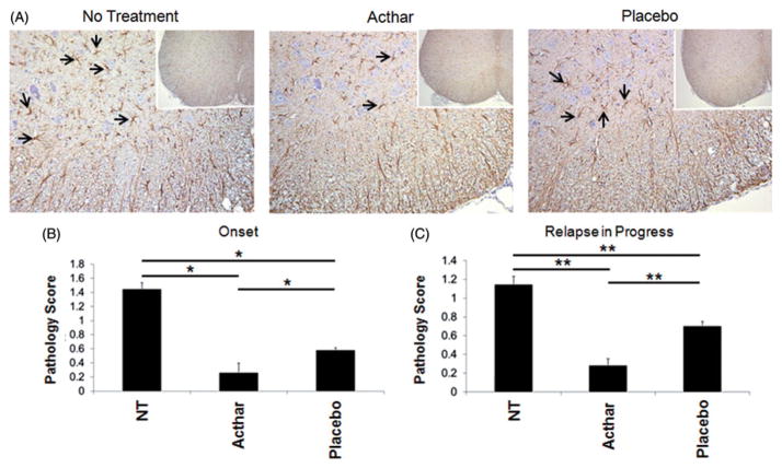
Figure 4.
Acthar dampens astrogliosis in the spinal cords of EAE mice. (A) Representative spinal cord tissue sections stained for GFAP, as described in the “Methods” section. Images shown at 20× magnification and insets at 10× magnification. (B) Quantification of GFAP scoring (described in the “Methods” section) of spinal cord tissue sections obtained from mice in which treatment was initiated at the onset of relapse (n = 15 for no treatment, 12 for Acthar treatment and 10 for Placebo gel treatment). *p<0.05, one-way ANOVA followed by Dunn’s test. (C) Quantification of GFAP scoring of spinal cord sections obtained from mice in which treatment was initiated after the onset of relapse (n = 16 for all groups of mice). **p<0.01, ANOVA followed by Holm–Sidak test. Values represent the mean ± SEM (at least six spinal cord tissue sections were scored for each mouse). NT no treatment.

