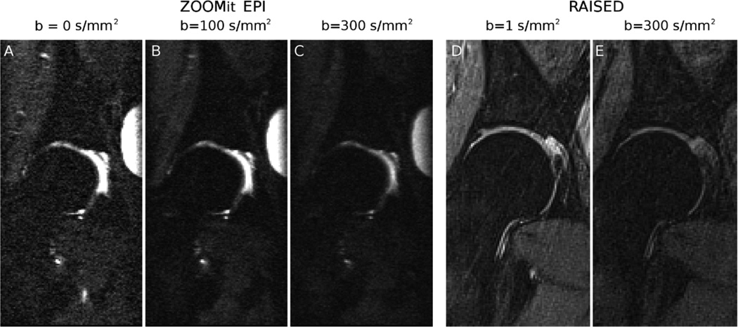
Figure 4.
Example of diffusion-weighted images of the femoral head on a 34-years old female asymptomatic subject. A–C. Imaging sequences include a DW-EPI acquired with the parallel transmit technology to selective excite a small region around the femur head (syngo ZOOMit; Siemens Healthcare, Erlangen, Germany). Parameter acquisition for the EPI were TE/TR = 71/6600 ms, slice thickness = 3.3 mm, echo-train length = 63, 13 slices, matrix size = 192×96 mm2, in-plane resolution = 0.80×0.80 mm2, bandwidth = 1024 Hz/pixel, b-value = 0,100,300 s/mm2 with averages of 1, 3, 5, partial Fourier = 5/8, acquisition time = 5:35 min. D–E. Diffusion-weighted images acquired on the same volunteer with a RAISED sequence (TE/TR = 38/1500 ms, slice thickness = 3 mm, 13 slices, FOV = 154×154 mm2, in-plane resolution = 0.80×0.80 mm2, b-value = 0,350 s/mm2, bandwidth = 290 Hz/pixel, spokes = 114 [2.64 acceleration with respect to the Niquist condition], acquisition time = 2:50 min per b-value).

