Figure 5.
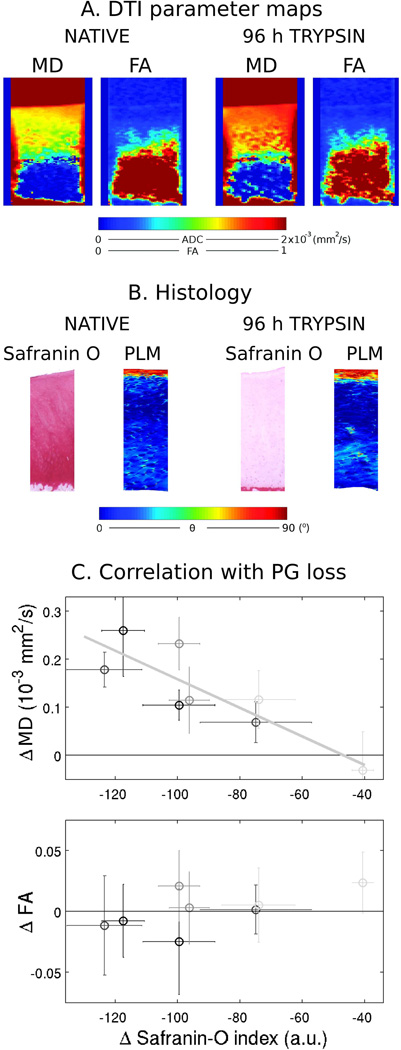
Change of diffusion parameters with progressive PG depletion. A. Diffusion parameter maps (MD and FA) of a sample before and after a 96 h treatment with a low dose (0.1 mg/ml) of trypsin for 96 hours. PG depletion resulted in increased MD but no change of FA. B. Histology analysis of the same sample before and after treatment with trypsin. Histology included safranin-O sensitive to the PG content and polarized light microscopy (PLM) for analysis of the collagen structure. Treatment with trypsin led to decrease in the intensity of the safranin-O staining, but no change in the collagen architecture. C. Correlation between the change in MD (r2 = −0.86, P<0.007) and FA (no significant) with the loss in PG content measured on safranin-O histology slides. Error bars represent the 2σ intervals of the difference to baseline. Light to dark gray encodes increasing trypsin incubation times (6, 48, 72 and 96 h). [Adapted from data of Raya et al. (24)].

