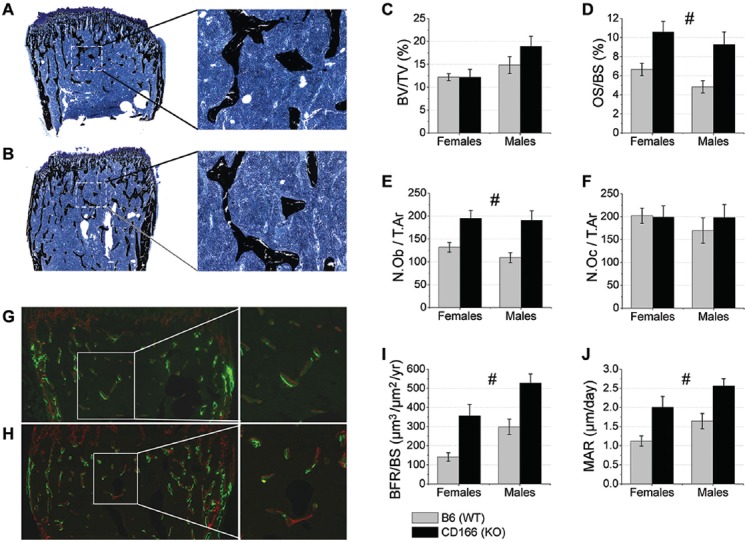
Figure 3.

Static and dynamic trabecular bone phenotype histomorphometric analysis of 6 week-old, WT and CD166-/- distal femurs. Trichrome staining of (A) WT and (B) CD166-/- distal femurs, original magnification, 10X (inset 20X). (C) BV/TV, (D) OS/BS (%), (E) N.Ob/T.Ar, and (F) N.Oc/T.Ar. Dual fluorochrome labeling of (G) WT and (H) CD166-/- distal femurs, original magnification 10X (inset 20X). (I) BFR/BS and (J) MAR. n=6-8 mice/group. Results are presented as the mean ± SE. #Indicates significant main effect for genotype, with no significant genotype and sex interaction (p<0.05).
