
Fig. 2.
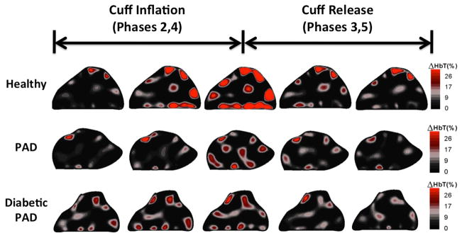
This figure shows coronal cross-sectional images from a healthy subject, PAD patient and diabetic PAD patient. The five frames shown outline the hemodynamics during the thigh cuff occlusion and release. The healthy volunteer has a greater amount of blood pooling in the leg during the occlusion as well as a faster reaction to the application and release of the thigh cuff.