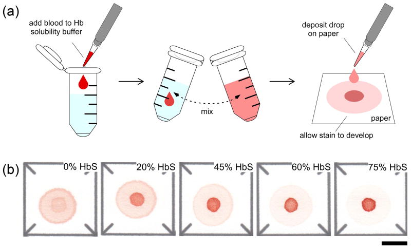
Figure 1.
Schematic illustration of the paper-based HbS assay and the characteristic blood stains. (a) To perform the assay a 20 μL droplet of blood mixed with Hb solubility buffer (phosphate buffer containing saponin and sodium hydrosulfite) is deposited on chromatography paper, a blood stain is allowed to form (polymerized deoxy-HbS is trapped in the paper mesh and soluble forms of Hb are wicked laterally), and the stain is scanned and analyzed automatically to estimate %HbS in the sample. (b) Characteristic blood stains produced on paper by samples with various %HbS. Scale bar is 1 cm.

