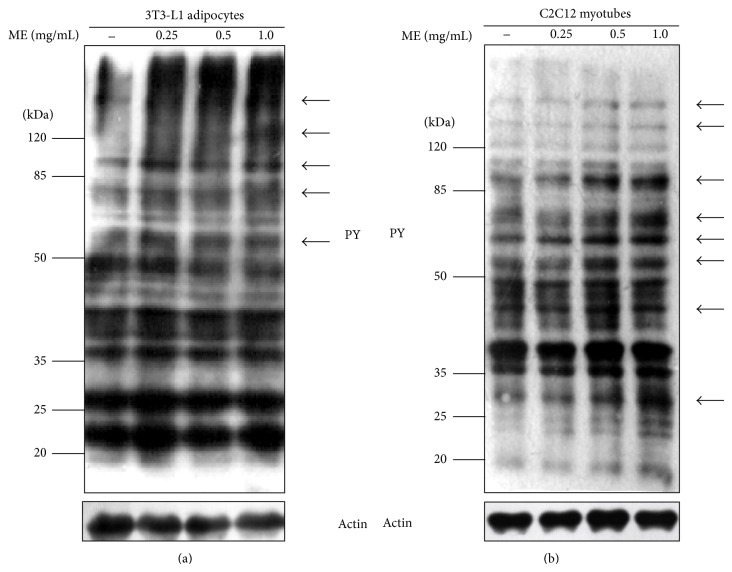
Figure 3.
Tyrosine phosphorylation of cellular proteins stimulated by ME. Differentiated 3T3-L1 adipocytes (a) and C2C12 myotubes (b) were stimulated with different doses of ME for 30 min. Equal amounts of total proteins were subjected to Western blot analysis with antibodies against phosphotyrosine (pY) and actin. Arrows indicate proteins whose phosphorylation is significantly enhanced by ME.

