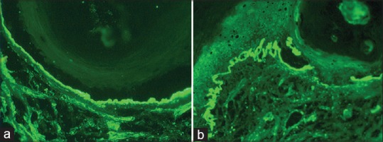
Figure 3.

Systemic lupus erythematosus (SLE) – (a) Direct immunofluorescence (DIF) showing dense granular continuous deposits of immunoglobulin G (IgG) (++++) along the dermo-epidermal junction (lupus band test) (fluorescein isothiocyanate anti-IgG, ×200) SLE – (b) DIF showing dense granular continuous deposits of IgA (++++) along the dermo-epidermal junction (lupus band test) (fluorescein isothiocyanate anti-IgA, ×200)
