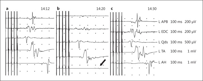
Fig. 2.
Patient 9. Reversible MEP deterioration in a 38-year-old man diagnosed with a right frontal parasagittal AVM based on partial motor seizures in the left leg. a Baseline traces corresponding to the TES MEP of the left upper and lower limb muscles during a second Onyx injection. b Focal change restricted to a decrease in MEP amplitude of >90% in the left AH muscle (black arrow). c After 10 min, embolization was terminated, and the MEP returned to its baseline level with no increase in the stimulation threshold. L = Left; EDC = extensor digitorum communi; Qds = quadriceps.

