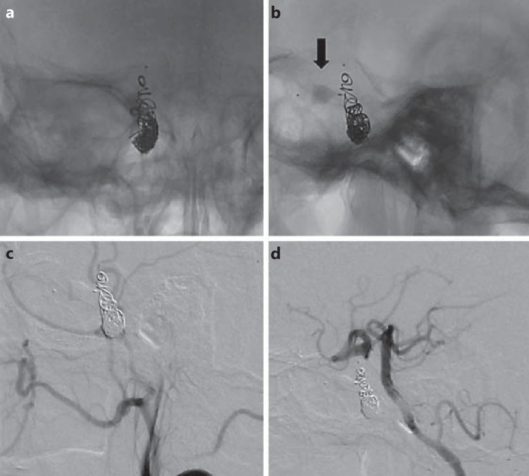
Fig. 4.
a, b Plain films in posteroanterior and lateral views showing the MVP (arrow) in the ICA distal to the fistula and coil mass in the ICA proximal to the fistula. c, d DSA in lateral views of the right common carotid and left vertebral artery showing persistent complete anterograde and retrograde obliteration of the high-flow CCF.

