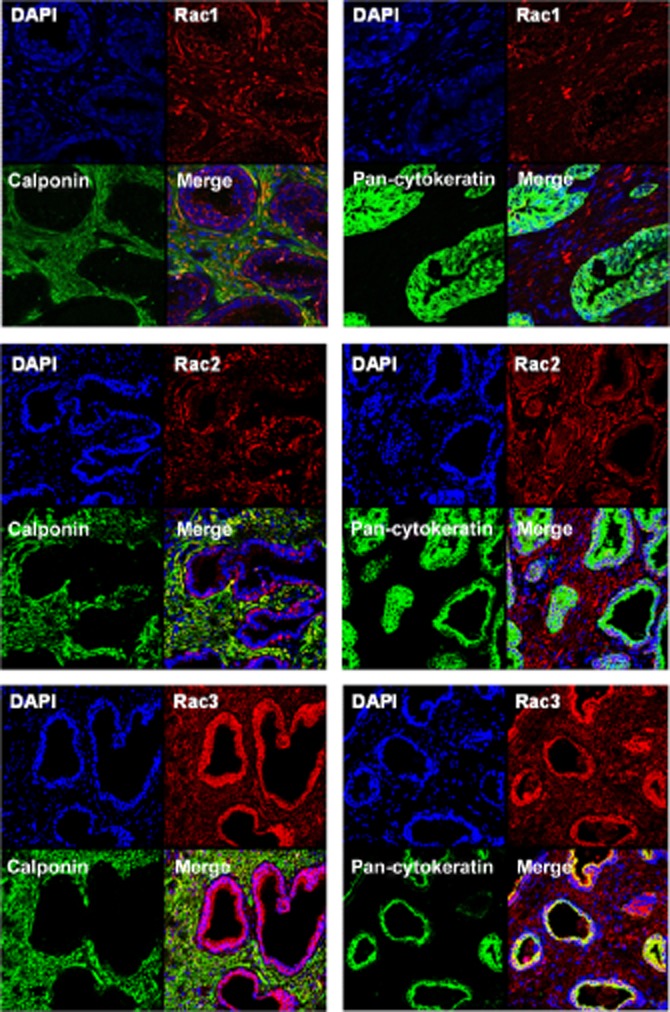
Figure 2.

Fluorescence staining of human prostate tissue. Tissues were double-labelled using isoform-specific antibodies for Rac isoforms 1–3, and calponin (left panels) or pan-cytokeratin (right panels). Yellow colour indicates colocalization of immunoreactivities. Shown are representative stainings from a series of tissues from n = 6 patients, with similar results.
