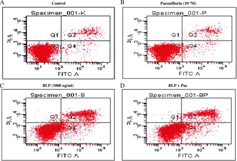
Fig. 3.
Effects of BLP and paeoniflorin on apoptosis of THP-1 cells. Control cells were treated with incomplete RPMI- 1640 medium (A). THP-1 cells were treated for 24 h with paeoniflorin 10–4 M (B), with 1,000 ng/ml BLP (C), or with BLP 1000 ng/ml + Pae 10–4 M (D). The cells were harvested for quantification of apoptosis by flow cytometry measured as the percentage of Annexin V-FITC positive cells

