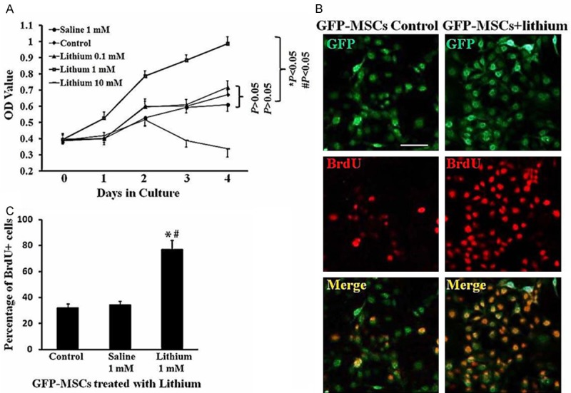
Figure 2.

Promotion of lithium on GFP-MSCs proliferation in vitro. A. Growth curves. Disassociated GFP-MSCs at P3 were plated in 96 well plates. The cells were exposed with lithium chloride at various concentrations (n = 6 wells for each) for 0, 1, 2, 3, 4 days. After various treatments, cell viability was determined by MTT assay. B. BrdU labeling. GFP-MSCs were treated with lithium (1 mM, n = 5 wells) or saline (1 mM, n = 5 wells) for 48 h. BrdU (10 mM) were added to each groups of GFP-MSCs, and incubated for 2 h, followed by immunostaining with BrdU antibody. Green: GFP; red: BrdU+. Scale Bar = 100 μm. C. Quantitative analysis of BrdU incorporation. The number of BrdU positive cells was shown as a percentage of the total numbers of GFP-positive cells, *P < 0.05, comparison between control GFP-MSCs and lithium-treated GFP-MSCs. #P < 0.05, comparison between saline-treated GFP-MSCs and lithium-treated GFP-MSCs.
