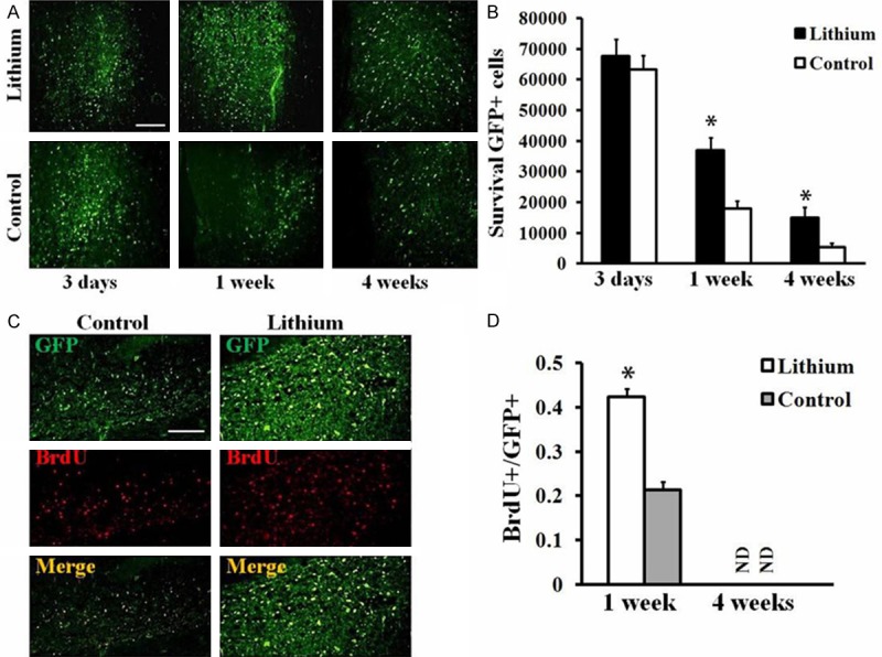
Figure 4.

Effects of lithium on GFP-MSCs survival and proliferation after transplantation into the spinal cord. A. Grafted GFP-MSCs in the spinal cord of lithium or saline (Control) treated animals. Spinal cord of rats in each group was harvested at day 3, 7 and 28 and sections were prepared. Transplanted GFP-MSCs were found in sections of spinal cord based on the expression of GFP (green). Scale Bar = 100 μm. B. Quantitative analysis of GFP-positive cells in sections of spinal cord. Data represent mean ± S.D., 5 sections per rat, n = 6 rats in each group, *P < 0.05, comparison between GFP-MSCs treated control and lithium plus GFP-MSCs treated animals. C. BrdU labeling. Spinal cord of rats in each group were harvested at 1 and 4 weeks post transplantation, and sections were prepared and immunostained with BrdU antibody. Increased BrdU+ (red)/GFP+ (green) cells were found in the spinal cord of rats treated with lithium by compared with control animals at 1 week after cell transplantation. D. Quantitative analysis of BrdU+/GFP+ cells of the sections from rat spinal cord at 1 week or 4 weeks post cell transplantation. Data represent mean ± S.D., 4 sections per rat, n = 6 rats in each group, *P < 0.05, comparison between GFP-MSCs treated control and lithium plus GFP-MSCs treated animals.
