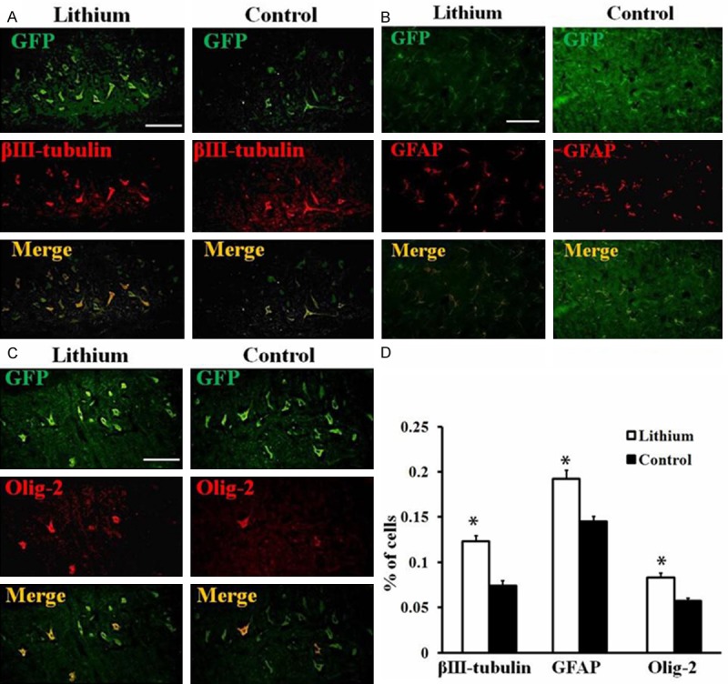
Figure 5.

Promotion of lithium on GFP-MSCs neural differentiation after transplantation into rat spinal cord. (A-C) Neural differentiation of grafted GFP-MSCs in rat spinal cord. Spinal cord of rats in each group was harvested at 4 weeks post transplantation and sections were prepared. Transplanted GFP-MSCs differentiated into (A) βIII-tubulin+ neurons, (B) GFAP+ astrocytes and (C) olig-2+ oligodendrocytes after 4 weeks post transplantation in the spinal cord. Scale Bar = 50 μm; (D) Quantitative analysis of percentages of GFP-MSCs differentiated into each type of neural cells among total numbers of GFP-positive cells. Data represent mean ± S.D., 4 sections per rat, n = 6 rats in each group, *P < 0.05, comparison between GFP-MSCs treated control and lithium plus GFP-MSCs treated animals.
