
Figure 2.
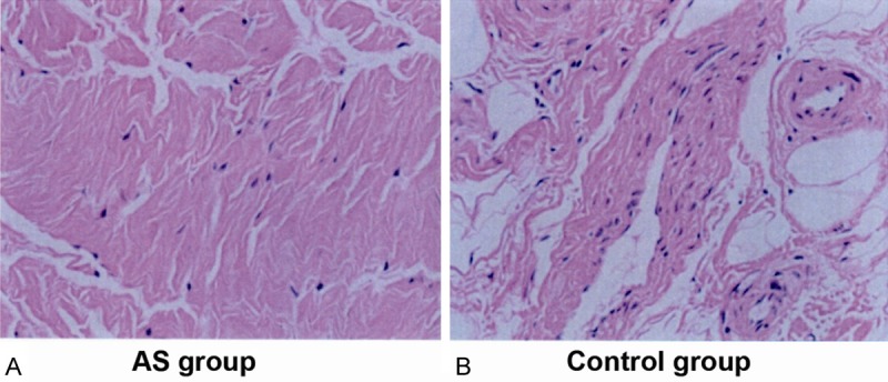
Hematoxylin-eosin (HE) staining of ligament tissue in (A) ankylosing spondylitis (AS) and (B) control group. The ligament tissue was mainly composed of longitudinal arrangement of elastic fibers (along the long axis), amongst which collagen fibers existed. The main ligament cells were scattered fibroblasts. The cell numbers in the control group appeared to be greater than those in the AS group by visual analysis.