
Figure 3.
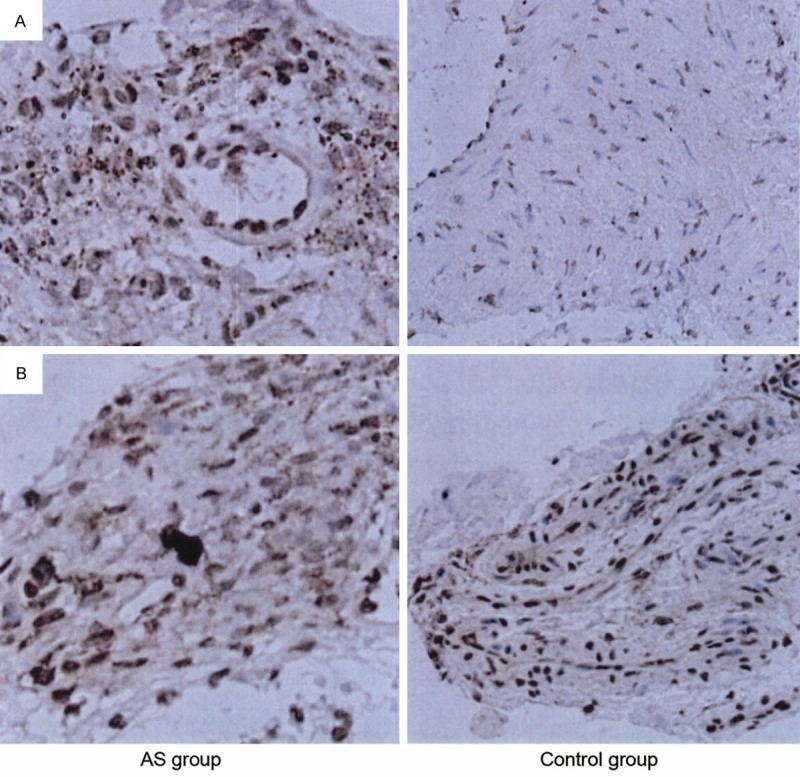
A. Immunohistochemistry staining of Notch1 in ankylosing spondylitis (AS) group and control group; B. Immunohistochemistry staining of hairy and enhancer of split (HES) in ankylosing spondylitis (AS) group and control group. The intense positive signals of Notch1 and Hes localized in the nucleus (yellow or brown particles). The expression of Notch1 and Hes in ligament tissue of the AS group increased significantly compared with control group.