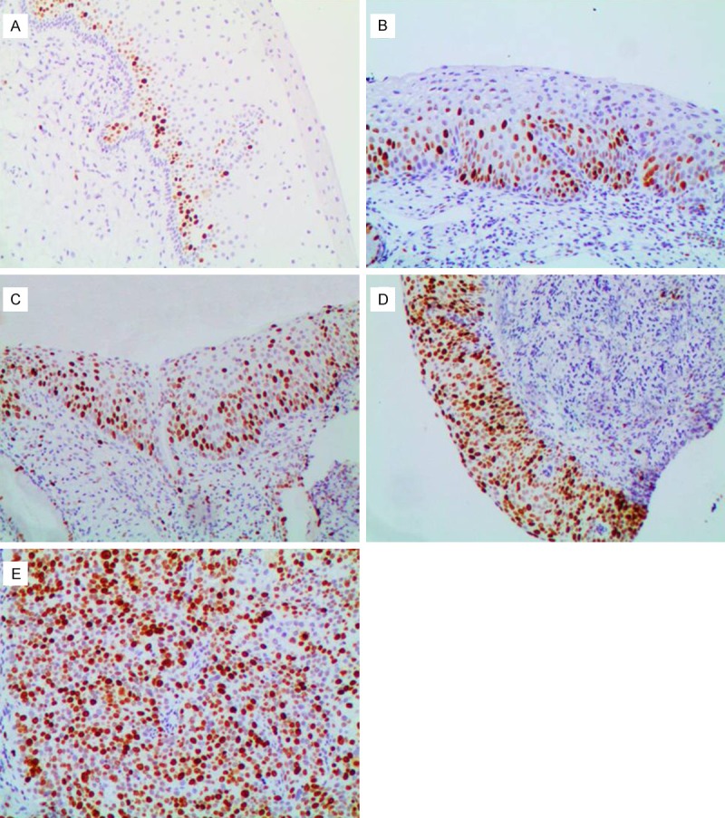
Figure 3.

Immunohistochemical staining of Ki-67 (100 ×). A: In negative for dysplasia, negative staining in normal cervical tissue except in the basal and parabasal cells; B: In cervical intraepithelial neoplasia1 (CIN1): diffuse Ki-67 immunostaining restricted to the lower third of the cervical epithelium; C: In CIN2, diffuse Ki-67 immunostaining of two-thirds of the cervical epithelium; D: In CIN3, diffuse Ki-67 immunostaining of full thickness positivity of the dysplastic epithelium; E: In squamous cell carcinoma, diffuse strong positive.
