
Figure 1.
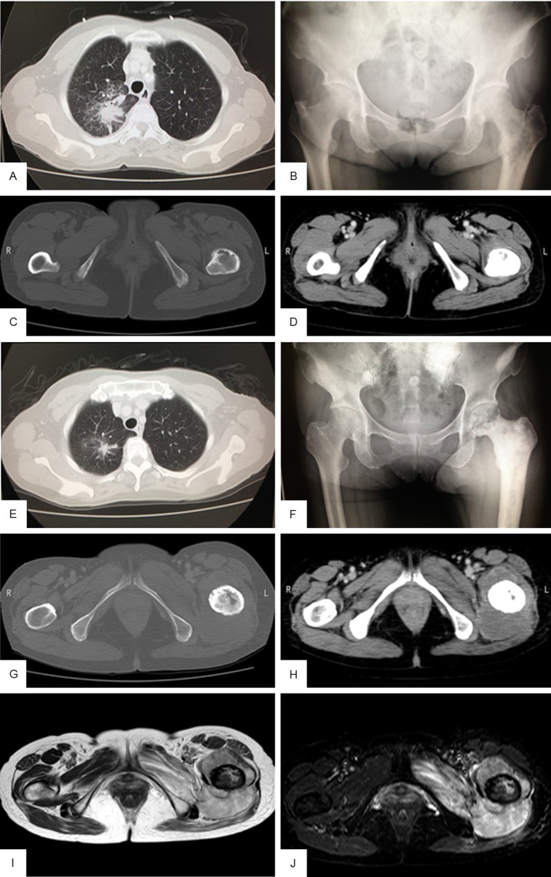
Presurgical imaging of the primary tumor and femoral metastasis. (A) Computed tomography (CT) of the chest showed a high-density nodular mass before gefitinib treatment. (B) Plain radiography revealed an osteolytic lesion in the left proximal femur before gefitinib treatment. (C, D) Contrast enhanced CT of the femurs and pelvis showed a thin cortex in the left femur with no soft tissue mass before gefitinib treatment. (E) Chest CT taken during gefitinib treatment and irradiation revealed marked shrinkage of the tumor. (F) Plain radiograph taken before surgery showed an osteosclerotic lesion with osteolytic change in the left proximal femur and decreased permeability into the surrounding soft tissue. (G, H) Enhanced CT taken before surgery revealed an isodense mass within the bone marrow and surrounding soft tissue. (I, J) Magnetic resonance imaging performed before surgery revealed a mass with isointensity on T2-weighted images (I) and with heterogenous intensity on fat-suppressed T2-weighted images (J) around the left femur.