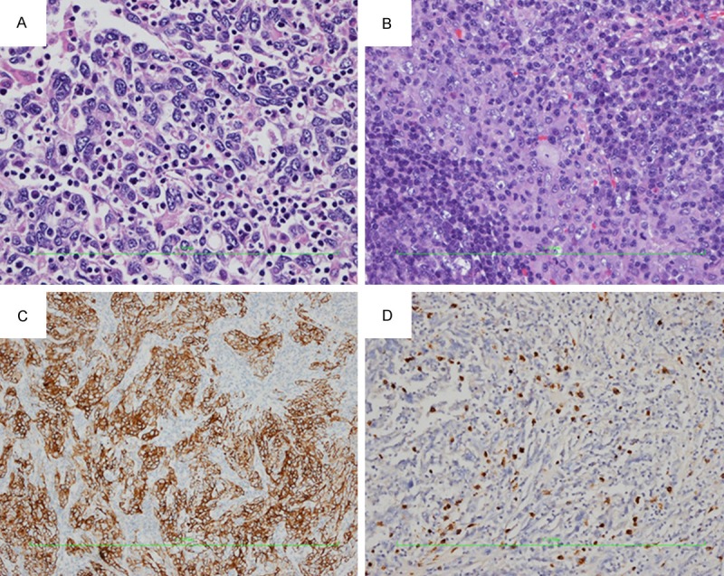
Figure 3.

A. Proliferation of atypical large cells, characterized by an eosinophilic cytoplasm, with large nuclei and prominent nucleoli. Epithelial cells were surrounded by a dense lymphoid stroma, extending inside the tumor (HE, ×400). B. Metastatic lymph node with poorly differentiated hepatocellular carcinoma in center (HE, ×200). C. Immunohistochemical staining for AE1/AE3 is (2+) positive in hepatocellular LELC (IHC, ×100). D. Immunohistochemical staining for EBER is negative in epithelial cells of hepatocellular LELC (IHC, ×100).
